use
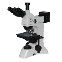
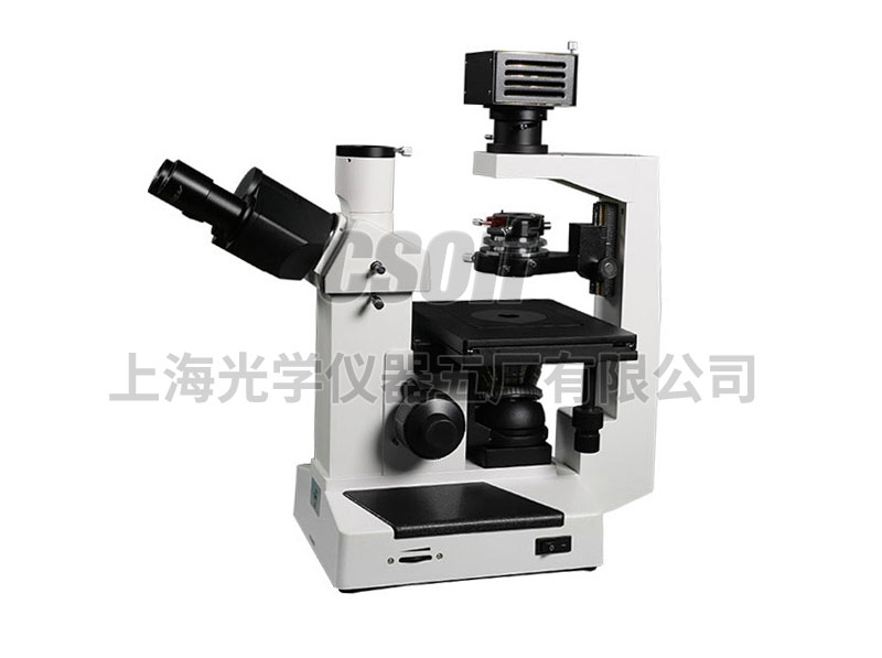
The instrument is equipped with a long working distance plan achromatic objective, a large field eyepiece and an adjustable condenser. The product has a compact structure and a reasonable operating part design, which makes the observer feel relaxed and comfortable during the operation of the instrument. The product is suitable for microscopic observation of cell tissue, transparent liquid tissue, and dynamic microscopic observation of cultured tissue in culture dishes. It can be used in scientific research institutes, colleges and universities, medical and health, inspection and quarantine, agriculture, animal husbandry and dairy industries.
The instrument can be equipped with a digital photography system, and the image is processed, edited, saved and output by the computer. The system can also be further connected to a microscopic image analysis system to perform metrological detection, morphological analysis, research statistics and output graphic reports on microscopic images. Covering almost all application fields of image quantitative analysis, all kinds of detection and analysis related to image morphology can be completed.
Specification
| category | Magnification/Field of View Diameter (mm) | configure |
| big vision flat field eyepiece |
WF10×/20 | have |
| WF16×/13 | apolegamy | |
| WF20×/11 | apolegamy |
| Class No | magnification times | numerical aperture(NA) | cover-glass thickness(mm) | working distance(mm) | Phase contrast ring plate |
| Long working distance plan achromatic Objective PLL |
10× | 0.25 | 1.2 | 8.8 | have |
| 25× | 0.4 | 1.2 | 4.8 | apolegamy | |
| 40× | 0.6 | 1.2 | 3.3 | apolegamy |
Optional system
Image inverted microscope 37X-V configuration
1. Host: Inverted Biological Microscope 37XC
2. Dedicated Image Adapters
3. Digital camera system (USB interface)
4. Computer and printer (bring your own or optional)


