Introduction
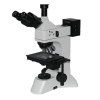
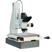
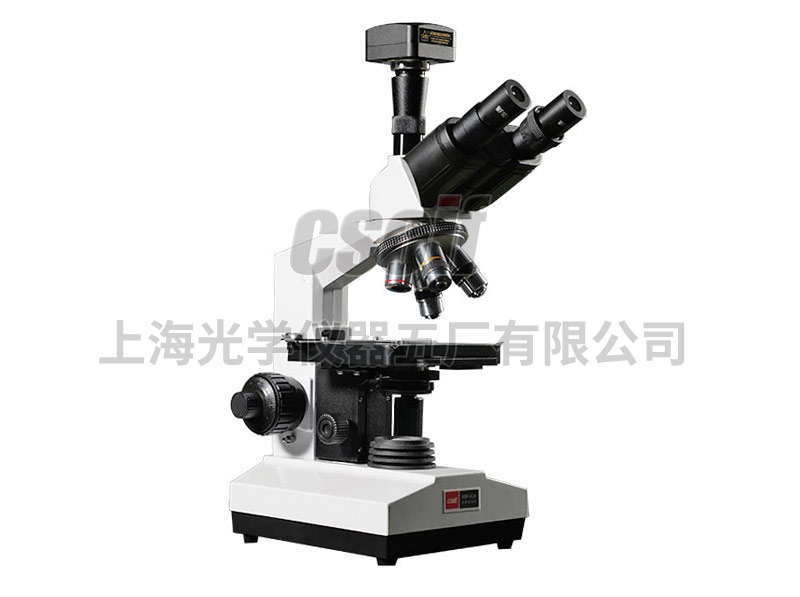
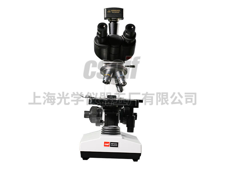
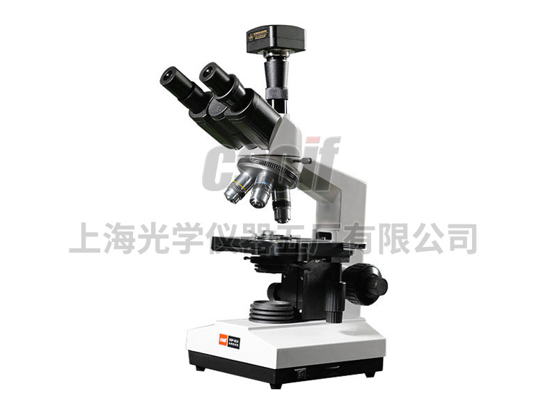
Image microscope is a conventional microscopic imaging, which is enlarged and displayed on a video monitor through a high-definition CCD camera, which is convenient for observation and research; a digital camera can also be used. Digital photography of images.
Then the image is processed, edited, saved and output (such as printing, etc.) or entered into the multimedia system and electronic mailbox by the microcomputer.
The optional microscopic image analysis system can further process and analyze the image, and all kinds of detection, analysis and statistics related to image morphology, such as: tissue and cell morphology; oil, water, blood component content; microparticles , line pattern and various geometric quantities of detection, analysis, statistics, etc., can be completed.
Technical Specifications
1. Trinocular Tube: Hinged 360° Positioning, 30° Inclination
Interpupillary distance and diopter adjustable
2. Total magnification: 40×~1600× (visual);
120×~3000×(image)
3. Mechanical barrel length: 160㎜
4. Worktable: 160×140㎜ Movement range: Horizontal 76㎜ Vertical 50㎜ Vernier grid value: 0.1㎜
5. Focusing mechanism: Coaxial coarse and fine adjustment Focusing limit device Adjustment range: 35㎜ Fine adjustment value: 0.002㎜
6. Abbe Condenser: Numerical aperture NA1.25 with variable aperture and color filter Can be raised and lowered structure Adjustment range: 40mm
7. Lighting system: LED lighting system
8. Power supply: 110V or 220V 50Hz
9. Object, eyepiece and magnification: (in the table: total magnification condition of image system: 1/3′ CCD 14′ display screen)
| flat field eyepiece | Achromatic Objectives | system | Field of view diameter
(㎜) |
working distance
(㎜) |
total magnification | ||
| Magnification / Field of View Diameter (mm) | Magnification | Numerical aperture | microscope | image system | |||
| 10×/18
16×/13 |
4× | 0.10 | 干 | 4.5 | 37.5 | 40×/64× | ≈120× |
| 10× | 0.25 | 1.8 | 7.31 | 100×/160× | ≈300× | ||
| 40× | 0.65 | 0.45 | 0.63 | 400×/640× | ≈1200× | ||
| 100× | 1.25 | 油 | 0.17 | 0.18 | 1000×/1600× | ≈3000× | |
System Configuration
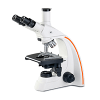
1. Biological microscope: XSP-8CA main unit (trinocular)
2. digital camera system
3. Lenovo business computers and HP color inkjet printers: (bring your own or purchase)


