Time:2022-03-28 Click:751
(1) Appropriate thickness. This thickness enables high resolution in better optics and is compatible with the system's depth of field.
(2) Transparent enough.
(3) Sufficient contrast can be formed on the basis of absorption difference.
Almost all biological materials can be obtained by paraffin embedding, sectioning (or smearing), pasting, paraffin dissolution, clearing, etc. to meet the requirements of specimen thickness and transparency, and then a certain degree of contrast can be obtained by staining procedures.
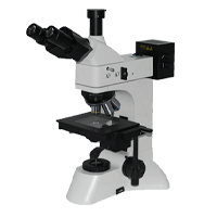
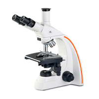
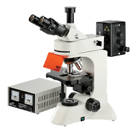
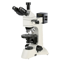
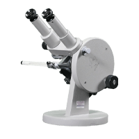
Biological and medical tissue sections commonly used by Olympus transmission light microscopes are usually 3-7 microns thick. In this thickness range, when viewed with a low power objective (eg, 10x), the depth of field is fully sufficient to observe the entire slice thickness; when viewed with a high magnification objective (eg, 100x), the depth of field is only a fraction of the slice thickness. Constant fine-tuning from top to bottom can give an overall impression of a slice. Therefore, a conventional section with a thickness of 5^-7um can be considered as a compromise between the ideal of low magnification and high magnification, both of which allow for better depth of field and resolution. At the same time, due to various reasons, this harmonic thickness also meets many requirements in practical work. , the fine structure of objects is not always first; second, in very thin (1-V25m) slices, the three-dimensional relationships of biological specimens are not visible. Conversely, there is a mutual benefit at different levels of the biological sample, in addition to a discernible loss of resolution, when sections of 8 to 10 um or greater thickness are used.
Specimens of animal and plant material are sufficiently thin and transparent after fixation, embedding, sectioning, paraffin dissolution and clarification
But the contrast is small, mainly because of refraction and diffraction. Clearly, this specimen is far from ideal by comparison. But this covers it with an "absorber" by reducing the effect of refraction, and increases the contrast through the difference in absorption. Refraction between the specimen and the surrounding area can be reduced by wrapping the specimen in Canadian gum or other synthetic wrapping medium. When all of these encapsulated media are hardened by evaporation or polymerization, their refractive indices are close to those of the major constituents of fixed or dehydrated plant and animal tissue. In most animal and plant tissues, the value is between 1.51 and 1.54. This method also eliminates glare caused by reflections under the coverslip.
For more than a century, an effective method has been staining, which contrasts well with Olympus microscope specimens. In general, unstained biological samples, especially when placed in a medium with a refractive index similar to the biological sample, have little contrast in the images. In dyed samples, the increase in contrast is based on the selective distribution of the dye. The contrast between stained and unstained specimens is very different.
Biosections are usually attached to glass slides with standard dimensions of 26-76 mm and thickness of 1.1-1.3 mm. The sides of the slide should be parallel planes. The requirements for glass carrier thickness are not very critical, but when the thickness exceeds the free working distance of some highly corrected concentrators, the glass carrier thickness becomes important for focusing the light source and creating the field aperture in the object plane. In this case, glass slides with a thickness of 0.9^-1.0 mm are suitable.


Whatsapp:Jason Fa