Time:2022-03-28 Click:918
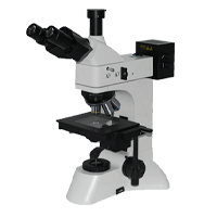
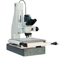
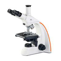
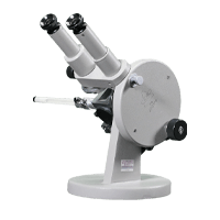
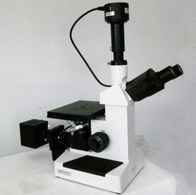
Electron microscopes and optical microscopes are optical instruments that are widely used in many fields. Electron microscope is a large-scale instrument that takes the electron beam as the illumination source, transmits or reflects the sample through the electron flow and multi-stage magnification of the electromagnetic lens, and then images on the fluorescent screen. An optical microscope is an optical instrument that uses visible light illumination to form an enlarged image of tiny objects. The main differences between electron microscopes and optical microscopes are as follows:
1. Different lighting sources. The illumination source of electron microscope is the electron flow emitted by electron gun, and the illumination source of light mirror is visible light (old light or lamp). Because the wave length of electron flow is much shorter than the wavelength of light wave, the magnification and resolution of electron microscope are significantly higher than that of light mirror.
2, the lens is different. The objective lens that magnifies in the electron microscope is an electromagnetic lens (an annular electromagnetic coil that can generate a magnetic field in the middle), while the objective lens of the optical mirror is an optical lens made of glass. There are three groups of electromagnetic lenses in the electron microscope, which are equivalent to the condenser, objective and eyepiece in the light microscope.
3. The imaging principle is different. In the electron microscope, the electron beam acting on the sample to be inspected is magnified by an electromagnetic lens and then hits a fluorescent screen for imaging or acts on a photosensitive film for imaging. The mechanism of the difference in the density of the particles is that when the electron beam acts on the sample to be tested, the incident electrons collide with the atoms of the substance to produce scattering. Since different parts of the sample have different scattering degrees for the electrons, the electron image of the sample is presented in shades. The object image of the sample in the light microscope is presented with a difference in brightness, which is formed by the difference in the amount of light absorbed by the different structures of the sample, which is also the key to the difference between the electron microscope and the optical microscope.
4. The preparation methods of the specimens used are different. The preparation procedure of tissue and cell specimens used for electron microscope observation is complicated, and the technical difficulty and cost are high. Special reagents and operations are required in the steps of sampling, fixation, dehydration and embedding. Put the embedded tissue block into the ultrathin microtome and cut it into ultrathin specimens with a thickness of 50 ~ 100nm. Specimens observed under light microscope are usually placed on glass slides, such as ordinary tissue slice specimens, cell smear specimens, tissue pressing specimens and cell drop specimens.

Whatsapp:Jason Fa